ads/wkwkland.txt
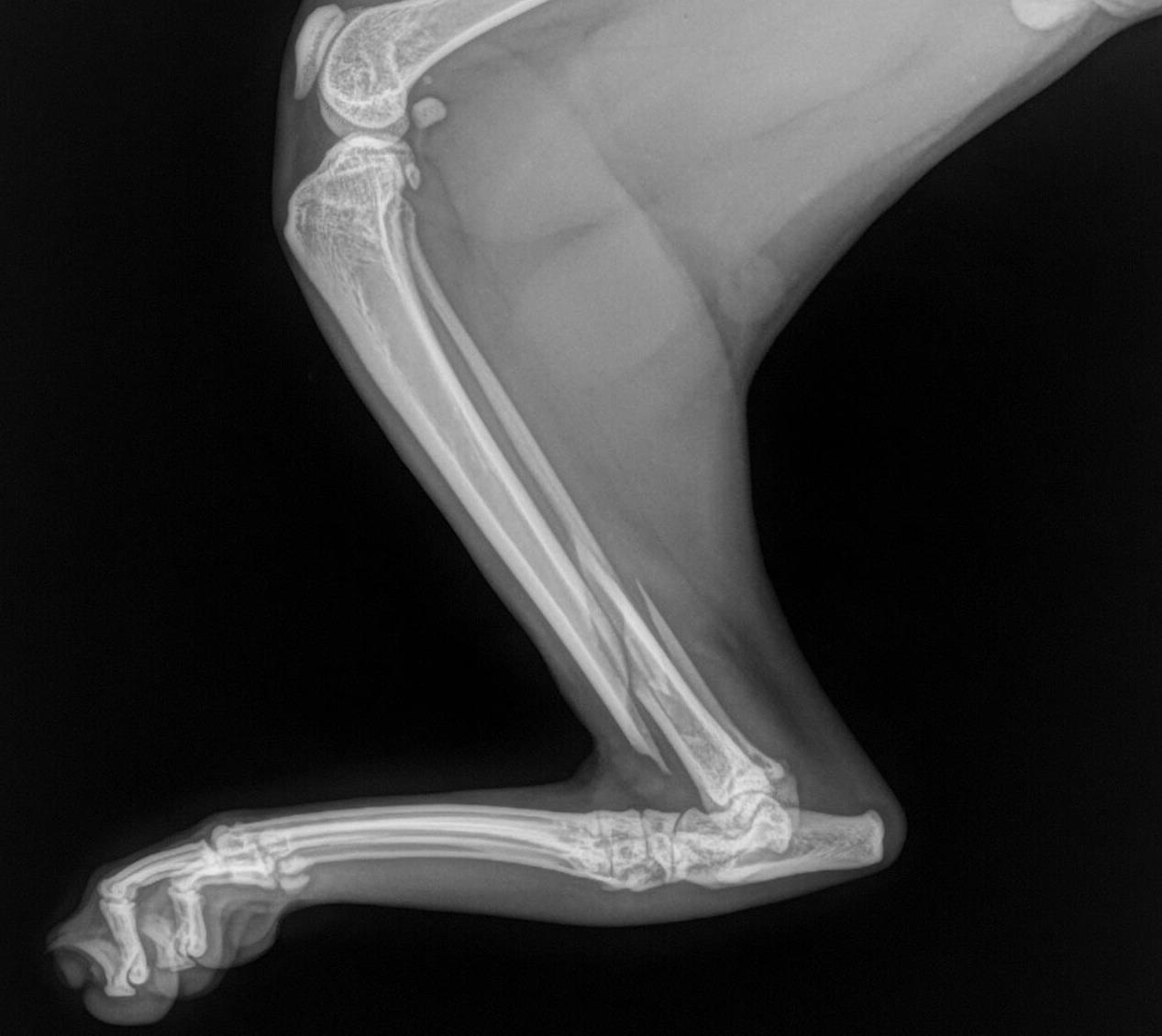
47 Best Pictures Cat X Ray Anatomy : Normal chest x-ray: Anatomy tutorial | Kenhub. Anatomy xray of the pelvis. How to read a chest xray, see chest xray atlas, yale lung anatomy, pic #1, #2, #3, #4, #5 cat scan anatomy: Learn about x ray anatomy with free interactive flashcards. This is a radiograph of the abdomen of a normal cat that is laying. The humeral head should be located at the level of the glenoid.
ads/bitcoin1.txt
Learn about x ray anatomy with free interactive flashcards. Learn about cat x ray costs and important facts about them. See more ideas about radiology, cat scan, respiratory therapy. I find them really difficult because their sleek bodies hide the anatomy. Anatomy xray of the pelvis.

As the pace of veterinary advancement accelerates, even the most experienced veterinary teams are.
ads/bitcoin2.txt
The humeral head should be located at the level of the glenoid. 10:10 top viral talent recommended for you. Anatomy xray of the pelvis. Buy 4d vision cat anatomy model: Learn about cat x ray costs and important facts about them. Normal anatomy in an axial image of the shoulder (a/b). This is a radiograph of the abdomen of a normal cat that is laying. It is important to know what to expect and differences between. Ct #1 , ct #2 , ct #3, ct #4, ct#5, ct#6, ct#7 and more ct here ct. As the pace of veterinary advancement accelerates, even the most experienced veterinary teams are. Each of these anatomical structures should be viewed using a systematic approach. After that we will do some radiograph reading lessons, teaching you about the normal anatomy of dogs and cats. 8:12 vaank recommended for you.
I find them really difficult because their sleek bodies hide the anatomy. It is important to know what to expect and differences between. Learn about cat x ray costs and important facts about them. The humeral head should be located at the level of the glenoid. Domestic cat studies drawn from a skeleton in the taxidermy lab (which we affectionately call the dead room) at the university of newcastle.
Buy 4d vision cat anatomy model:
ads/bitcoin2.txt
I find them really difficult because their sleek bodies hide the anatomy. I draw a lot of big cats but almost never house cats. 4.7 out of 5 stars 73 ratings. Tissues, computed tomography (ct) imaging, also known as cat scanning (computerized axial. Each of these anatomical structures should be viewed using a systematic approach. As the pace of veterinary advancement accelerates, even the most experienced veterinary teams are. But if your cat needs veterinary x rays, the amount of radiation she'll be exposed to is relatively small, and generally not excessive enough to cause any sort of health problem. The humeral head should be located at the level of the glenoid. If all other factors that affect radiog… Anatomy xray of the pelvis. After that we will do some radiograph reading lessons, teaching you about the normal anatomy of dogs and cats. Domestic cat studies drawn from a skeleton in the taxidermy lab (which we affectionately call the dead room) at the university of newcastle. Each of them can be distinguished body, front and back ends.
It is important to know what to expect and differences between. Domestic cat studies drawn from a skeleton in the taxidermy lab (which we affectionately call the dead room) at the university of newcastle. Learn about x ray anatomy with free interactive flashcards. After that we will do some radiograph reading lessons, teaching you about the normal anatomy of dogs and cats. Ct #1 , ct #2 , ct #3, ct #4, ct#5, ct#6, ct#7 and more ct here ct.

I find them really difficult because their sleek bodies hide the anatomy.
ads/bitcoin2.txt
This is a radiograph of the abdomen of a normal cat that is laying. Facsimile reproduction of a copy held by the amnh library. The humeral head should be located at the level of the glenoid. 4.7 out of 5 stars 73 ratings. After that we will do some radiograph reading lessons, teaching you about the normal anatomy of dogs and cats. I find them really difficult because their sleek bodies hide the anatomy. 10:10 top viral talent recommended for you. Why do cats need them? Domestic cat studies drawn from a skeleton in the taxidermy lab (which we affectionately call the dead room) at the university of newcastle. Those branches are acynonyx, panthera, and felis. It is important to know what to expect and differences between. Each of these anatomical structures should be viewed using a systematic approach. Learn about x ray anatomy with free interactive flashcards.
ads/bitcoin3.txt
ads/bitcoin4.txt
ads/bitcoin5.txt
ads/wkwkland.txt

0 Response to "47 Best Pictures Cat X Ray Anatomy : Normal chest x-ray: Anatomy tutorial | Kenhub"
Post a Comment